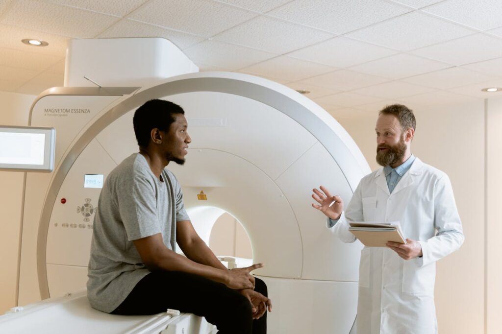
PET-CT, or Positron Emission Tomography-Computed Tomography, is an advanced medical imaging technique that combines two powerful imaging modalities to provide detailed anatomical and functional information about the body. Here’s an in-depth exploration of PET-CT:
- Principle: PET-CT integrates Positron Emission Tomography (PET) and Computed Tomography (CT) technologies into a single imaging session. PET detects positrons emitted by a radiopharmaceutical tracer, while CT provides detailed anatomical images using X-rays.
- Radiopharmaceuticals: Patients receive a radiopharmaceutical tracer, typically fluorodeoxyglucose (FDG), which is injected intravenously and accumulates in tissues based on metabolic activity. FDG is particularly useful for imaging areas with high glucose metabolism, such as tumors.
- PET Imaging: Positrons emitted by the tracer interact with electrons in the body, producing gamma rays. Detectors surrounding the patient capture these gamma rays to create images that show metabolic activity and molecular processes within tissues.
- CT Imaging: Simultaneously, the CT component takes X-ray images from multiple angles. These images provide detailed structural information, such as the size, shape, and location of abnormalities detected by PET.
- Combined Images: PET-CT combines functional PET images showing metabolic activity with anatomical CT images, allowing for precise localization and characterization of abnormalities, such as tumors or areas of inflammation.
- Applications: PET-CT is used for:
- Oncology: Staging and monitoring of cancers, assessing response to treatment, and detecting cancer recurrence.
- Cardiology: Evaluating myocardial perfusion, detecting coronary artery disease, and assessing cardiac function.
- Neurology: Mapping brain activity, diagnosing neurodegenerative diseases, and evaluating epilepsy.
- Infection and Inflammation: Detecting infections and inflammatory conditions throughout the body.
- Advantages: PET-CT provides comprehensive information in a single imaging session, improving diagnostic accuracy and treatment planning. It helps in distinguishing between benign and malignant lesions, guiding biopsy and surgical procedures.
- Patient Experience: The PET-CT procedure is non-invasive and generally well-tolerated. Patients may need to fast before the scan, and some may receive contrast dye for enhanced CT imaging.
- Safety Considerations: The radiation dose from PET-CT scans is carefully controlled and optimized for diagnostic purposes. The benefits of the information gained from the scan outweigh the risks of radiation exposure.
- Technological Advancements: Ongoing research focuses on improving image resolution, developing new radiopharmaceutical tracers for specific diseases, and integrating PET-CT with other imaging modalities for enhanced diagnostic capabilities.
PET-CT imaging represents a significant advancement in medical diagnostics, offering clinicians valuable insights into disease processes and guiding personalized treatment strategies based on individual patient needs.


