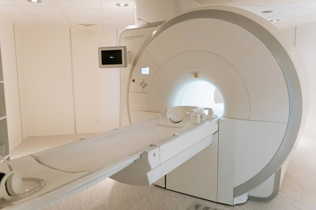
MRI, or Magnetic Resonance Imaging, is a powerful medical imaging technique that uses strong magnets and radio waves to create detailed images of organs and tissues inside the body. Let’s delve into the fascinating world of MRI:
- Principle: MRI utilizes a strong magnetic field and radiofrequency pulses to create detailed images of organs, tissues, and structures within the body.
- Types of MRI: Different types include:
- T1-weighted MRI: Provides detailed anatomical images with excellent tissue contrast.
- T2-weighted MRI: Highlights differences in water content of tissues, useful for detecting abnormalities like edema.
- Functional MRI (fMRI): Measures brain activity by detecting changes in blood flow.
- Diffusion MRI: Maps the diffusion of water molecules in tissues, aiding in stroke diagnosis and assessing fiber tracts in the brain.
- Uses: MRI is used to diagnose and monitor a wide range of conditions, including:
- Neurological Disorders: Brain and spinal cord tumors, strokes, multiple sclerosis.
- Musculoskeletal Issues: Joint injuries, spinal disorders, soft tissue tumors.
- Cardiovascular Conditions: Heart anatomy, blood flow patterns, and abnormalities.
- Abdominal and Pelvic Disorders: Liver and kidney diseases, tumors, and reproductive system conditions.
- Procedure: During an MRI scan, the patient lies inside a large, tube-like machine. The MRI machine generates a magnetic field and radio waves, which create detailed cross-sectional images of the body. The procedure is painless but may be noisy due to the MRI machine’s magnetic coils.
- Contrast Agents: Some MRI scans may require contrast agents (gadolinium-based) injected into the bloodstream to enhance the visibility of certain tissues or blood vessels.
- Safety: MRI does not use ionizing radiation, making it safer than techniques like CT scans. However, precautions are taken regarding metal implants, pacemakers, and claustrophobia.
- Image Interpretation: Radiologists interpret MRI images to diagnose conditions based on tissue density, structure, and contrast characteristics. Advanced software aids in image analysis and 3D reconstruction.
- Advantages: MRI provides high-resolution images of soft tissues and does not expose patients to ionizing radiation, making it ideal for repeated imaging and pediatric patients.
- Limitations: MRI scans can be time-consuming, and patients must remain still during the procedure to avoid image blurring. Certain metallic implants or foreign bodies may interfere with imaging quality.
- Future Directions: Ongoing research focuses on improving MRI technology, developing faster imaging sequences, enhancing image resolution, and exploring new clinical applications.
MRI continues to revolutionize medical diagnostics, offering precise anatomical and functional information that aids in early diagnosis, treatment planning, and monitoring of various medical conditions.


